Beranda
/ Tendon Diagram Hand / Hand Anatomy Eorthopod Com / Tendons are made up of tough bundle of fibrous tissue, which connects muscles and bones.
Tendon Diagram Hand / Hand Anatomy Eorthopod Com / Tendons are made up of tough bundle of fibrous tissue, which connects muscles and bones.
Insurance Gas/Electricity Loans Mortgage Attorney Lawyer Donate Conference Call Degree Credit Treatment Software Classes Recovery Trading Rehab Hosting Transfer Cord Blood Claim compensation mesothelioma mesothelioma attorney Houston car accident lawyer moreno valley can you sue a doctor for wrong diagnosis doctorate in security top online doctoral programs in business educational leadership doctoral programs online car accident doctor atlanta car accident doctor atlanta accident attorney rancho Cucamonga truck accident attorney san Antonio ONLINE BUSINESS DEGREE PROGRAMS ACCREDITED online accredited psychology degree masters degree in human resources online public administration masters degree online bitcoin merchant account bitcoin merchant services compare car insurance auto insurance troy mi seo explanation digital marketing degree floridaseo company fitness showrooms stamfordct how to work more efficiently seowordpress tips meaning of seo what is an seo what does an seo do what seo stands for best seotips google seo advice seo steps, The secure cloud-based platform for smart service delivery. Safelink is used by legal, professional and financial services to protect sensitive information, accelerate business processes and increase productivity. Use Safelink to collaborate securely with clients, colleagues and external parties. Safelink has a menu of workspace types with advanced features for dispute resolution, running deals and customised client portal creation. All data is encrypted (at rest and in transit and you retain your own encryption keys. Our titan security framework ensures your data is secure and you even have the option to choose your own data location from Channel Islands, London (UK), Dublin (EU), Australia.
Tendon Diagram Hand / Hand Anatomy Eorthopod Com / Tendons are made up of tough bundle of fibrous tissue, which connects muscles and bones.. Fortunately, there are exercises you can perform to strengthen and increase flexibility in your finger tendons. The epb tendon, along with the apl, also takes the thumb away from the hand. Each finger has two flexor tendons, the flexor digitorum superficialis (fds) and the flexor digitorum profundus (fdp). Closed tendon injuries of the hand and wrist in the athlete. They connect to 5 metacarpal bones that form the palm of the hand.
Inflammation of the tendon sheath results in trigger finger. The red lines show where the tendons attach the muscles to the bones. Tight tendons in your fingers can inhibit your ability to perform your daily routine. They are controlled by muscles in the forearm. Tendons in your fingers may become tight as the result of overuse injuries, such as tendinitis, or traumatic injuries, including fracturing a finger.
Racgp Hands Fingers Thumbs Assessment And Management Of Common Hand Injuries In General Practice from www.racgp.org.au Profundus tendons, which pass through the palm side of the wrist and hand, and attach at the bases of the. The tendons work like long ropes connecting the muscles of the forearm with the bones of the fingers and thumb. The peroneal tendons run down together behind the outer side of the ankle and then split before attaching to different parts of the foot. The tendons that allow each finger joint to straighten are called the extensor tendons. The epb tendon, along with the apl, also takes the thumb away from the hand. They act with the profundus tendons to flex the wrist and mcp and pip joints. The flexor tendons of the hand and fingers pass through the carpal tunnel of the wrist before separating in the palm and moving toward their ultimate attachment sites in the fingers. Superficialis tendons, which pass through the palm side of the wrist and hand, and attach at the bases of the middle phalanges.
Digits that extend from the palm of the hand, the fingers make it possible for humans to grip the smallest of objects.;
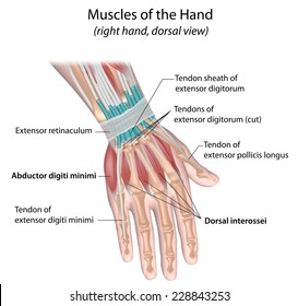
It is inflamed in the conditioned called de quervain's syndrome. The tendons that allow each finger joint to straighten are called the extensor tendons. Extensor tendons connect to muscles in the middle of the forearm, then extend through the wrist and hand to each finger, where they form the extensor hood. The radial and ulnar collateral ligaments are important to provide stability of the fingertip during pinching. Various methods of attaching the posterior deltoid to the. There are 3 major types of bones in the hand itself, including: Tendons allow fingers to pinch, grasp, grip, and straighten. Superficialis tendons, which pass through the palm side of the wrist and hand, and attach at the bases of the middle phalanges. Originates from the upper part of the fibula, passes underneath the foot and attaches by the medial foot arch peroneus brevis: The red lines show where the tendons attach the muscles to the bones. This condition typically causes pain and swelling, and it may result in a reduced range of movement in the. A hand strain is a stretching or tearing of fibers in muscles or tendons, the tissue that anchors muscle to bone. Hand a hand is a prehensile multi fingered appendage located at the end of the forearm or forelimb of primates such as humans chimpanzees monkeys and lemurs human anatomy for the artist the dorsal hand the dorsal the easiest tendons to identify in the dorsal hand are those of the extensor digitorum muscle its name means extensor of the digits which is
The main tendons of the hand are: The hand is composed of many different bones, muscles, and ligaments that allow for a large amount of movement and dexterity. Interventions for treating mallet finger injuries. Strains often occur in tendons that connect the muscles of the forearm to bones in the fingers. It is inflamed in the conditioned called de quervain's syndrome.
Flexor Tendon Repair And Rehabilitation from assets.aboutkidshealth.ca Tendons are thick, fibrous cords that connect muscles to bones. Related posts of thumb flexor tendon anatomy shoulder bones anatomy diagram. Digits that extend from the palm of the hand, the fingers make it possible for humans to grip the smallest of objects.; Tendons allow fingers to pinch, grasp, grip, and straighten. Examples that help finger movement include: Tendons are made up of tough bundle of fibrous tissue, which connects muscles and bones. There are 3 major types of bones in the hand itself, including: The fds inserts on the midportion of the.
The radial and ulnar collateral ligaments are important to provide stability of the fingertip during pinching.
Tendons of the hand are thin, flat and white in color. They are controlled by muscles in the forearm. The epb tendon, along with the apl, also takes the thumb away from the hand. We hope this picture hand nerve, tendon, and muscle anatomy can help you study and research. Inflammation of the tendon sheath results in trigger finger. Each finger has two flexor tendons, the flexor digitorum superficialis (fds) and the flexor digitorum profundus (fdp). When fingers joints straighten, they are being pulled by the extensor tendons. Fortunately, there are exercises you can perform to strengthen and increase flexibility in your finger tendons. Tendons are thick, fibrous cords that connect muscles to bones. Closed tendon injuries of the hand and wrist in the athlete. Trigger finger or trigger thumb, medically known as stenosing tenosynovitis, involves the pulleys and tendons in the hand that bend the fingers (see diagram). Reconstruction of the extensor carpi ulnaris (ecu. In the finger, the pulleys form a tunnel under which the tendons must glide.
Interventions for treating mallet finger injuries. When fingers joints straighten, they are being pulled by the extensor tendons. These wrist bones are attached to the radius and ulna of the forearm to form the wrist joint. If your extensor tendons are damaged, you'll be unable to straighten one or more fingers. Diagram of human forearm bones anatomy 10 photos of the diagram of human forearm bones anatomy anatomy elbow bones, anatomy shoulder bones, hand bones anatomy, human anatomy arm bones, leg bones anatomy, two bones in the forearm, wrist bones anatomy, hand, anatomy elbow bones, anatomy shoulder bones.
Hand Tendons Images Stock Photos Vectors Shutterstock from image.shutterstock.com Functional enthesitis at the extensor tendons of the toes. It is inflamed in the conditioned called de quervain's syndrome. Many tendons in your hands and feet attach to hand and foot bones, and help you move your fingers and toes. The terminal extensor tendon in the thumb comes from the extensor pollicis longus muscle. Tendonitis is an inflammation of the tendon. These tendons are used to either straighten or bend your fingers and thumb. Superficialis tendons, which pass through the palm side of the wrist and hand, and attach at the bases of the middle phalanges. Tight tendons in your fingers can inhibit your ability to perform your daily routine.
Hand a hand is a prehensile multi fingered appendage located at the end of the forearm or forelimb of primates such as humans chimpanzees monkeys and lemurs human anatomy for the artist the dorsal hand the dorsal the easiest tendons to identify in the dorsal hand are those of the extensor digitorum muscle its name means extensor of the digits which is
Tendonitis of the hand causes severe pain with movement of the hand. The radial and ulnar collateral ligaments are important to provide stability of the fingertip during pinching. They connect to 5 metacarpal bones that form the palm of the hand. They are controlled by muscles in the forearm. The epb tendon, along with the apl, also takes the thumb away from the hand. Shoulder bones anatomy diagram 9 photos of the shoulder bones anatomy diagram acromion anatomy, ankle bones anatomy, bones of the clavicle, elbow bones anatomy, hip bones anatomy, shoulder muscles anatomy, shoulder pain anatomy, hand, acromion anatomy, ankle bones anatomy, bones of the clavicle, elbow bones. If your extensor tendons are damaged, you'll be unable to straighten one or more fingers. Originates from the upper part of the fibula, passes underneath the foot and attaches by the medial foot arch peroneus brevis: Each finger has two flexor tendons, the flexor digitorum superficialis (fds) and the flexor digitorum profundus (fdp). Trigger finger or trigger thumb, medically known as stenosing tenosynovitis, involves the pulleys and tendons in the hand that bend the fingers (see diagram). All the complexity and demands can lead to an array of conditions that can make your hands hurt. The hand can be considered in four segments: We think this is the most useful anatomy picture that.
Tendon diagram of wrist : tendon diagram. The epb tendon, along with the apl, also takes the thumb away from the hand.